Thanks to NPR, I was directed to an op-ed that appeared in the New York Times in October 2012 entitled "In Cancer Care, Cost Matters." The article describes what may be one of the earliest accounts where doctors at Sloan-Kettering decided not to use a new cancer drug because of its extra cost when they were already using equally-effective drugs for their cancer treatments. The article piqued my interest. These days for any doctor who uses expensive technologies, it is easy to see where this evaluation will lead us in our new era of cost concerns in medicine. Medical device companies should take notice.
Few use more expensive technology in medicine than cardiologists and cardiac electrophysiologists. Not only is our technology expensive, it is also used frequently. Cardiovascular disease remains one of the largest cost drivers in medicine.
In the past, doctors (myself included) were complicit with device companies in our use of new technologies, implanting the latest model of defibrillator, for instance, not because it saved lives any better, but more so we could boast that our patients were receiving a "Cadillac" defibrillator rather than a "Ford" device (no disrespect intended to car companies, but you get my drift). After all, our patients deserved the best and most innovative technology available at the time. We did not want to be caught in the embarrassing position of being behind innovation power curve either.
I should acknowledge that there have been some very important recent developments with defibrillators since the 2005-2006 device recalls that plagued our specialty. The ability to monitor device reliability wirelessly facilitated the ability to detect device battery depletions or lead failures and has improved our understanding of tolerable device and lead failure rates, for instance. But it is not uncommon for a newly-named pacemaker or defibrillator to add several thousand dollars to the health care system over last year's device yet they may add only minor advances over the prior year's model.
Pacemakers, too, are being manufactured that are FDA-labelled "MRI-tolerant." But examples of safe application of MRI's to conventional pacemakers are prevalent in the medical literature, so is the extra cost of an MRI-safe pacemaker worth it? (In actuality, if you ask the informed pacemaker implanters out there, the main reason MRI pacemakers are routinely implanted is not because of safety concerns, but because of the CMS coverage decision for MRI payment in patients with pacemakers.(pdf)) How do we weigh the cost of these extra advances in technology versus their ability to improve our patients' actual morbidity and mortality?
Increasingly, doctors will soon be involved with purchasing decisions for advanced technologies. Pacemakers, defibrillators, implantable monitors, stents, catheters, and even small sheaths will be scrutinized for their utility, ease of use, and cost. (The liability of NOT using a technology will also be considered, but this issue is hard to measure and less familiar to frontline doctors.)
The real question, of course, is will the patient see these cost savings if older technologies are used? This is hard to say since there are so many layers to our health care system between the patient and the dollar. Also, how will patients react when they find out online that their model of medical device is last year's model rather than this year's? Will they seek another hospital system with shinier walls and bigger names? It's impossible to tell.
But if hospitals are smart, they'll make it clear to the patient on their bill what they saved themselves by using an earlier-year device. Medical device companies, too, might shift their marketing tactics to costs and benefits from things like size or shape of their device.
But one thing's for certain in the days ahead: device companies will have to carry not just this year's model of device, but last year's, too. Innovations in technologies will be harder to sell unless they show real patient morbidity or mortality benefit. Companies will have to adapt their marketing campaigns since cost savings, done correctly, will benefit everyone.
After all, it's now about real costs and benefits in health care, rather than just sexy bells and whistles.
-Wes
Addendum: The CMS National Coverage Decision for MRIs in patients with pacemakers is now functional.
Showing posts with label MRI. Show all posts
Showing posts with label MRI. Show all posts
Thursday, March 28, 2013


Friday, February 18, 2011
What's the Difference Between MRI-Safe and Conventional Pacemaker Leads?
... a little thicker, certainly, but otherwise (at least on the surface), not too much:
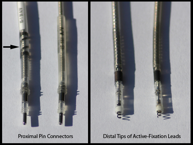
The recently-approved MRI-safe active-fixation lead from Medtronic (left lead in each frame) is compared to their conventional active-fixation lead. The arrow denotes the radio-opaque marker that can been seen on x-ray to identify the type of lead in the patient's body. A fluoro image of the two leads is shown below, again with Medtronic's MRI-safe lead on the left:
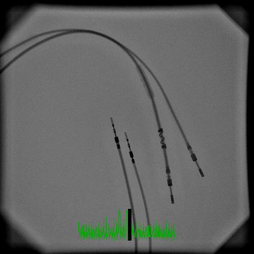
While the engineering hurdles were no-doubt considerable to make an MRI-safe pacemaker lead, given the growing body of evidence that newer pacemakers (when carefully monitored) can be scanned in MRI machines, I suspect the biggest difference in these leads is not their design per se, but rather the regulatory paperwork (and research) that had to be completed to document their safety.
Of course, the fact that CMS would not pay for MRI scans performed on patients with pacemakers before the advent of these newer devices probably also limited the number of scans performed.
-Wes
The recently-approved MRI-safe active-fixation lead from Medtronic (left lead in each frame) is compared to their conventional active-fixation lead. The arrow denotes the radio-opaque marker that can been seen on x-ray to identify the type of lead in the patient's body. A fluoro image of the two leads is shown below, again with Medtronic's MRI-safe lead on the left:
While the engineering hurdles were no-doubt considerable to make an MRI-safe pacemaker lead, given the growing body of evidence that newer pacemakers (when carefully monitored) can be scanned in MRI machines, I suspect the biggest difference in these leads is not their design per se, but rather the regulatory paperwork (and research) that had to be completed to document their safety.
Of course, the fact that CMS would not pay for MRI scans performed on patients with pacemakers before the advent of these newer devices probably also limited the number of scans performed.
-Wes
Wednesday, February 16, 2011
MRI-Safe Pacemakers - Version 1.0
Mary Knudson, a health journalist and author of the Heart Sense blog, does a great job covering the story behind the story on the newly approved MRI-safe pacemakers in a guest blog post at the Scientific American. She discusses the challenges ahead in regard to the widespread clinical adoption of MRI-safe pacemakers, the issues with Medicare coverage of MRI's of patients with these devices, the logistics involved in their use, and includes commentary from a number of physicians, including a tidbit from yours truly.
-Wes
-Wes
Monday, September 06, 2010
Take A Trip Down a Normal Right Coronary Artery
... courtesy of some cool video reconstruction:
UsefulProgress, a Paris-based startup, uses NVIDIA GPUs and CUDA technology to create stunningly detailed 3D stereo visuals like these from image sources such as X-ray, CRT and MRI.-Wes
Saturday, April 17, 2010
Heart Strings
Thanks to the wonders of magnetic resonance imaging (MRI), amazing images of the muscle fiber orientation of the left ventricle have been obtained:

-Wes
The image was produced using a branch of magnetic resonance imaging (MRI) called diffusion tensor imaging (DTI). The technique tracks the diffusion of water throughout the myocardium (the heart’s muscular wall comprising interconnected sheets of muscle cells called myocytes). Due to the way the myocytes are organized, the movement of water is restricted, so tracking the location of water molecules can reveal valuable information about the structure of the heart in a non-invasive way.Nice.
-Wes
Monday, November 17, 2008
Friday, February 01, 2008
Imaging Turf Wars
Well the inaugural issue of JACC Cardiovascular Imaging was just come out and heralds cardiologists as cardiovascular imaging experts. Eugene Braunwald's welcome letter was telling:
Why?
It's all about the money.
-Wes
"Despite its great advantages, selective angiography is an invasive technique that may be uncomfortable, entails some minimal risk to the patient, and is expensive. It can be applied only a limited number of times in each patient and it is not suitable for screening. Therefore, physicians caring for patients with known or suspected heart disease yearned for noninvasive cardiac imaging. Fortunately, 2 new techniques—echocardiography and nuclear imaging—became available in the 1960s and have improved progressively since then. Literally tens of millions of these examinations are performed each year, transforming medical practice. In the 1980s, 3 new noninvasive imaging techniques—computed tomography (CT), cardiac magnetic resonance (CMR) imaging, and positron emission tomography (PET, a nuclear technique)—emerged. Each of these provides information on cardiac structure and function that is an order of magnitude greater than the earlier imaging techniques.Radiologists are none too happy, I'm sure. No doubt they might take issue with the claim of "We need a new generation of cardiac imagers who are expert with the entire portfolio of modalities and who can provide a nonbiased selection of the technique that can best solve the clinical or research problem at hand." The friendly collaboration between radiologists and cardiologists went out the window long ago.
None of the aforementioned techniques are static and all are undergoing progressive refinement. Many important advances have been made by subspecialists in a single technique such as echocardiographers, nuclear cardiologists, and specialists in CT, CMR, or PET imaging, respectively. Each of these imaging modalities have their individual training programs, and the advances in these fields are described in specialized journals, each devoted to a single technique. Although this approach has been responsible for spectacular advances in each modality, it has led to fragmentation of patient evaluation, at times to competition between individual modalities, and to increased costs at a time of diminishing resources to pay for health care.
There is a growing consensus that the time has come to consider cardiovascular imaging in a more comprehensive, unified manner. We need a new generation of cardiac imagers who are expert with the entire portfolio of modalities and who can provide a nonbiased selection of the technique that can best solve the clinical or research problem at hand. To develop these broadly-based experts, we need new unified training programs that break down what are sometimes artificial interdepartmental and intradepartmental barriers.
Another important tool in shifting the imaging paradigm is to develop a journal of cardiovascular imaging. The Publications Committee of the American College of Cardiology and its talented staff, working closely with the College leadership, developed a vision for and then the concrete plans for the creation of JACC: Cardiovascular Imaging. The committee and its consultants selected the Editor-in-Chief, Dr. Jagat Narula, from a group of distinguished cardiac imagers. Dr. Narula has put together an outstanding group of associate editors, editorial board members, and consultants. They now deserve the cooperation of the entire cardiovascular community as they proceed on their important mission."
Why?
It's all about the money.
-Wes
Thursday, August 09, 2007
Medtronic Purchases MRI Patents
Medtronic made a necessary purchase of Biophan's intellectual property for its newly-introduced MRI-safe pacemaker product line. They got off pretty cheap: $11 million, and avoided later legal challenges that were sure to arise if they had not made the purchase. The next question is, since Biophan was best-known for their work in this space, will they have enough intellectual property left to survive?
-Wes
-Wes
Friday, July 20, 2007
Having Fun With MRI's
What do cardiology fellows and medical students to for fun? Heck, just throw the medical student in the MRI to look at his heart!
-Wes
Addendum: If you look closely, you can see a small central jet of tricuspid insufficiency during systole. (The tricuspid valve is the valve between the upper atrial chamber and the lower ventricular chamber on the left side of the movie. The dark flow of tricuspid insifficiency backward from the ventricle to the atrium is clearly seen. A small amount of this leakage is typically seen in normal hearts and is used to estimate the person's right heart pressures on echocardiography using the continuity equation.)
-Wes
Addendum: If you look closely, you can see a small central jet of tricuspid insufficiency during systole. (The tricuspid valve is the valve between the upper atrial chamber and the lower ventricular chamber on the left side of the movie. The dark flow of tricuspid insifficiency backward from the ventricle to the atrium is clearly seen. A small amount of this leakage is typically seen in normal hearts and is used to estimate the person's right heart pressures on echocardiography using the continuity equation.)
Friday, June 22, 2007
The MRI-Safe Pacemaker
Magnetic resonance imaging (MRI) scanners are one of the most effective ways to follow soft-tissue and vascular diseases in the body. Unfortunately, sometimes these patients also have heart disease that requires that a permanent pacemaker or implantable defibrillator be installed for control of heart rhythm abnormalities. For these patients, follow-up of their soft tissue tumors is impaired because MRI-safe pacemakers and defibrillators are not available on the market, especially for patients who are dependent on their pacemaker for their heart to beat at all. Fortunately, some investigators (in the US and Europe) have devised special protocols for non-pacemaker dependent patients to undergo MRI scans when they are clinically required. The FDA still frowns on this practice, however.
There are several issues with MRI's that can cause problems with conventional pacemaker and defibrillator systems:
Medgadget also has a an even better overview of Medtronic's study and the technology in place to counteract these issues.
I just hope that someday MRI-safe capabilities will eventually become a standard feature in all cardiac devices marketed. After all, patients may have their pacemakers for years before the need for an MRI arises.
-Wes
Studies presented by Sommer et al and Nazarian et al in this issue of Circulation offer further promising evidence in this regard. As with the previous studies, however, the authors acknowledge a multitude of limitations that prevent broad applicability of the results. Furthermore, one of these studies was not entirely free from concerning outcomes with the potential for serious clinical events. We view these results as consistent with our previous message that, on a case-by-case basis, the diagnostic benefit from MRI outweighs the presumed risks for some pacemaker and ICD patients. However, the FDA remains firm in its belief that those risks have not yet been characterized and mitigated sufficiently to justify the routine use of MRI in those populations.In February of this year, Medtronic began a trial overseas with a new MRI-safe pacemaker called Enrhythm MRI. The was trial recently announced in Canada as well. It will probably be a bit longer before we see the trial spread to the United States, although it looks like the centers have been selected that will participate.
There are several issues with MRI's that can cause problems with conventional pacemaker and defibrillator systems:
- Usually the most common is interference with sensing the patient's underlying rhythm (oversensing, usually of noise, causes the pacemaker to inhibit its output, or undersensing causes the pacemaker or defibrillator to not be able to see the underlying rhythm and may cause pacing at inappropriate times)
- Alteration in programming (like reverting to a power-on reset mode of pacing or actual damage to the circuitry)
- Changes in pacing thresholds, perhaps due to lead movement (torque) or local heating.
Medgadget also has a an even better overview of Medtronic's study and the technology in place to counteract these issues.
I just hope that someday MRI-safe capabilities will eventually become a standard feature in all cardiac devices marketed. After all, patients may have their pacemakers for years before the need for an MRI arises.
-Wes
Wednesday, January 17, 2007
Subscribe to:
Posts (Atom)