Showing posts with label EKG Du Jour. Show all posts

Tuesday, August 13, 2013

EKG Du Jour 32: The Misfiring Pacemaker

A dual chamber pacemaker was implanted the prior day by a local surgeon in the operating room.  The next morning, an EKG is obtained that showed the following:

Click to enlarge
You checked the CXR and all leads appeared to be in the proper location. 

Does the patient have to go back to the operating room?  Why or why not?

-Wes

Friday, May 03, 2013

EKG Du Jour #31: A Case of Chest Pain

A 61 year old patient presented to your ER with chest pain, low blood pressure, and this EKG:

(Click image to enlarge)

Now before you leap to the answer at the link provided below, ask yourself:

(a) What is this?
(b) What could cause this?
(c) How would you manage it?

And when you've really thought about it, click here for the answer but be prepared to describe what you see and what you'd do next.

-Wes

P.S.: (Yeah, I'm giving the answer now because it's Friday)

Saturday, September 08, 2012

EKG Du Jour #27: Caught in the Act

I've been looking at EKG's for over 20 years and I'm not sure I've ever seen this captured on a 12-lead electrocardiogam outside the EP laboratory. 

The patient was admitted with some vague chest discomfort.  The initial EKG and troponins were normal, but they decided to admit the patient anyway for observation.  Once on the ward after being given some heparin and aspirin, the patient was feeling perfectly fine - wanted to go home - but the second set of troponins returned positive.  The first year resident decided to order a second EKG. 

The EKG technician came dutifully, performed the EKG, and before she knew it, all hell broke loose:

Click image to enlarge

Fortunately, the patient did well after a quite bit of work in the cath lab.

Remember this story.  It might just save a life.

-Wes

Wednesday, February 01, 2012

EKG Du Jour #25 - The Exercise Enthusiast - Explained

Sorry about the delay, but unlike most of the EKG Du Jour series of EKG's, I elected to make a separate post to explain the interesting tracings that first appeared here.

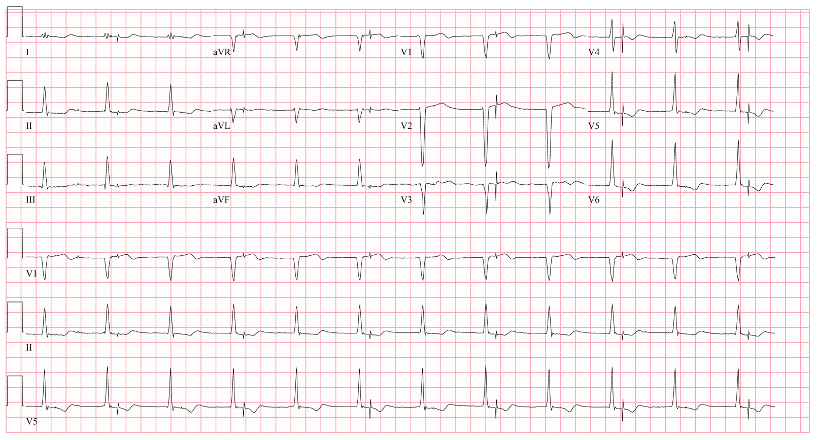
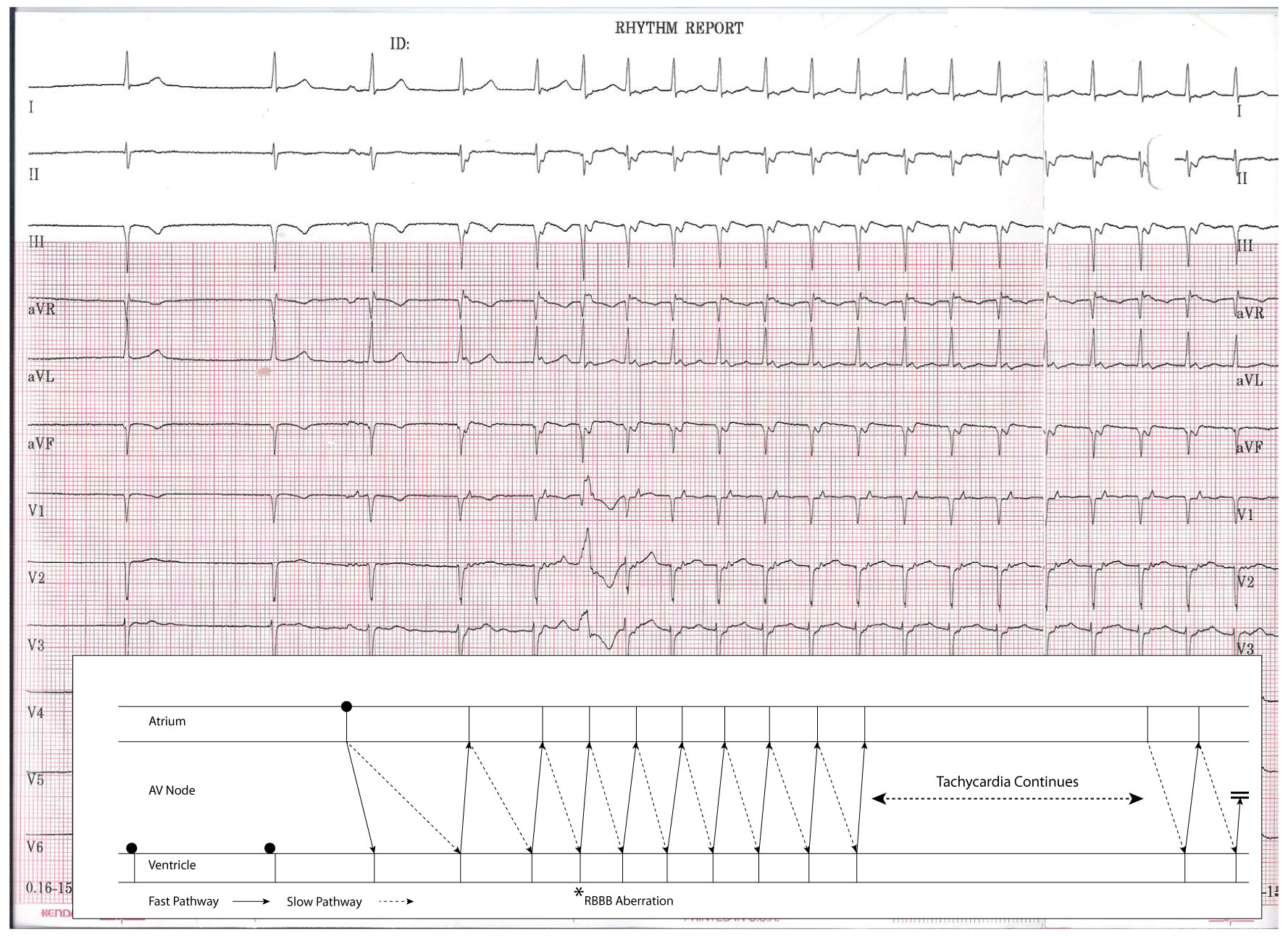
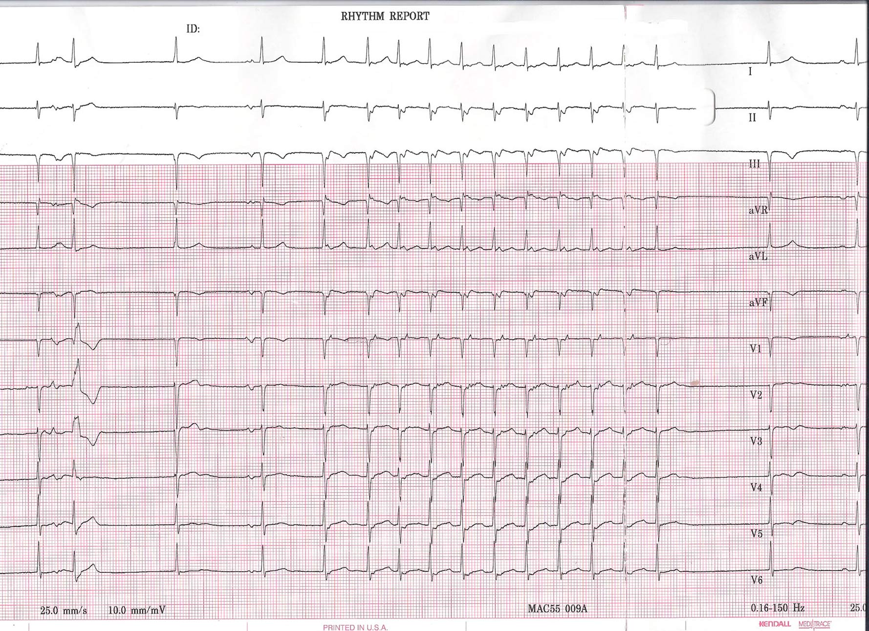
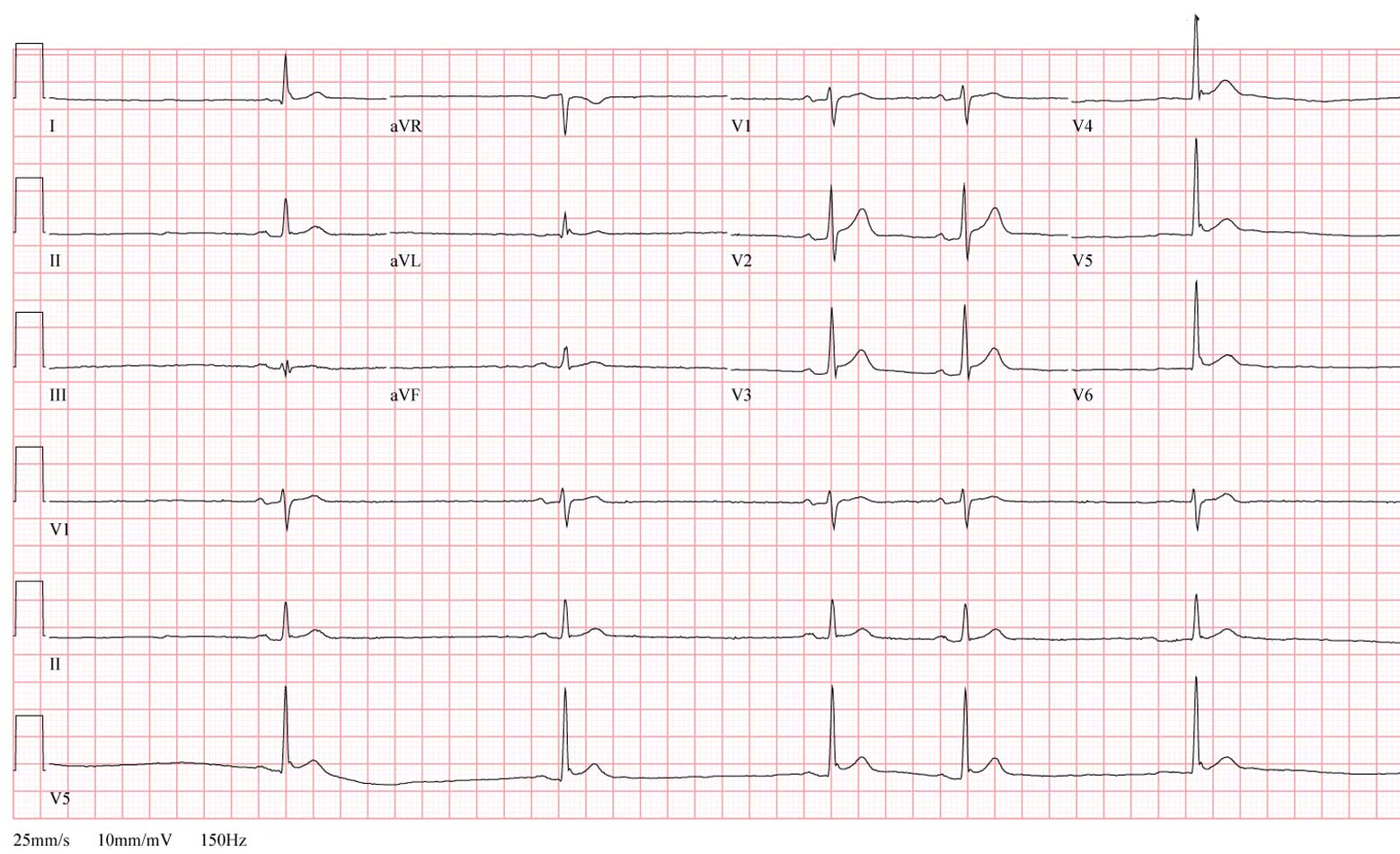
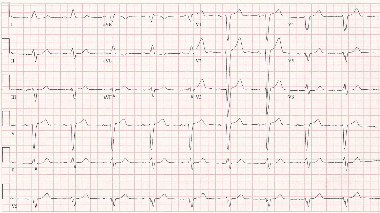
As a refresher, the original 12-lead rhythm strip obtained on this healthy, asymptomatic individual looked like this when he presented for evaluation of two near syncopal spells:

Click image to enlarge
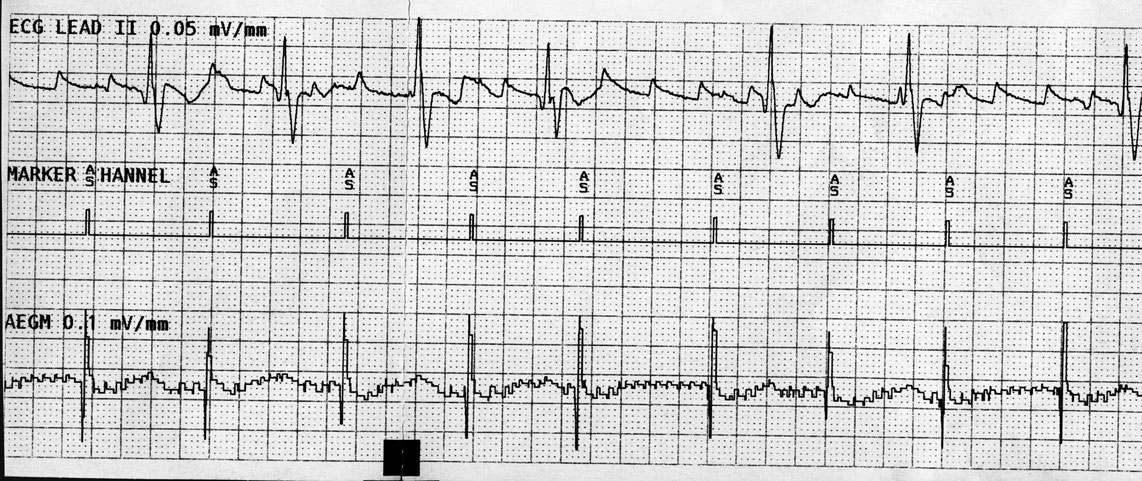
The first two beats do not appear to be preceded by a P wave but have a relatively narrow QRS complex associated with them, suggesting an AV junctional origin to the patient's rhythm. It is possible this man suffers from sick sinus syndrome or has a sinus rate that is slower than his junctional rate while at rest.

The third beat is where things get interesting. Here, a P wave is clearly seen that conducts to the ventricle with a normal PR interval. What is unsual, however, is the finding that the fourth beat (that appears to be junctional) occurs earlier than one would expect than a junctional escape beat to occur (based on the earlier two junctional beats). In effect, this fourth beat appears "pulled in" earlier to the preceding beat, but also has a clearly visible retrogradely-conducted P wave (best seen in V1) that occurs immediately after the QRS complex. At this point, the patient is in a supraventricular tachycardia that accelerates slightly with the fifth beat occurring earlier - probably because of hemodynamic alterations that occur due to the unusual cardiac activation sequence (slightly decreased BP and increased catecholamine level).

The sixth beat is a widened QRS complex of RBBB morphology. This beat is either a PVC (less likely) or (more likely) an aberrantly-conducted supraventricular beat. This beat aberrates because of the long-short nature of the initiation of the tachycardia that finds the right bundle branch refractory while the left bundle branch conducts to the ventricle. With continued tachycardia, the right bundle branch recovers and the tachycardia continues with a narrow QRS morphology.

The very last beat of the tracing defines the end of the run of supraventricular tachycardia.

So how did the tachycardia initiate?

This is an example of a normal P wave initiating SVT due to AV nodal "double-fire." That is, a single atrial beat conducts down BOTH the fast and slow pathway of the AV node. In this case, typical AV nodal reentrant tachycardia was initiated by a single sinus beat in this gentleman.

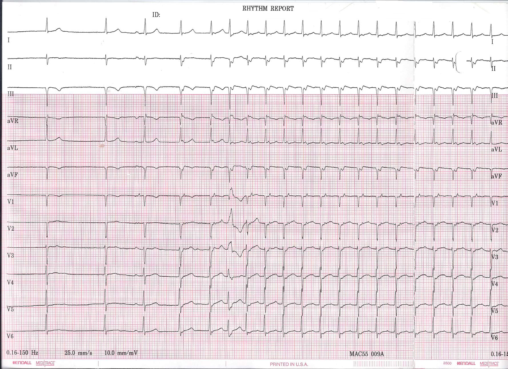
Here is the above tracing explained using a favorite of EP's -- a ladder diagram:

Click image to enlarge
To fix this problem, an EP study with slow pathway ablation should be performed with evaluation of the patient's residual sinus node function after the slow pathway is ablated. If his sinus node recover time remains sluggish after the ablation, then (and likely only then) should a permanent pacemaker be implanted.

-Wes

Thursday, January 26, 2012

EKG Du Jour #25 - The Exercise Enthusiast

He was a 65 year-old previously healthy avid exercise enthusiast on no medications who was referred for evaluation of two near-syncopal episodes. The first episode occurred approximately 10 minutes after he stopped exercising on a treadmill. He felt he had to grab the wall to prevent himself from falling.

The second occurred after riding his bike home from the gym. Just after riding his bike home, he became very lightheaded and felt he had to sit on his front porch and nearly lost consciousness. His symptoms passed after resting for about 20 minutes.

While having his blood pressure checked, the nurse noted an irregular pulse. He felt fine and was unaware of his heart rhythm. A 12-lead rhythm strip was obtained and two representative 12-lead rhythm strips from the recording are shown below:

Click image to enlarge


Click image to enlarge


Any ideas? What therapy (therapies?) would you recommend?

-Wes

Strips used with patient permission.

Monday, September 26, 2011

EKG Du Jour #24: Reversals

In the spirit of connecting via social media, this EKG Du Jour hails from the great Aussie blog, Life in the Fast Lane.

(There's lots to learn on many levels.)

-Wes

Thursday, September 22, 2011

EKG Du Jour #23 - With Increasing Prevalence: The Awful EKG

"Hey, Wes! I've been holding on to this EKG from a recent clinic visit with a patient of mine who presented with chest pain and thought it would make a cool EKG for your 'EKG Du Jour' series! I never could get a good tracing and I'm pretty sure my EKG machine works properly:"

Click image to enlarge


For now, forget the chest pain. What's the patient's principle diagnosis?

(Trust me: you'll be seeing more of this.)

-Wes

h/t: Gary Novak, MD

Clue #1: Still uncertain? A major hint to the answer is here, so look only if you must.

Clue #2: By the way, this is not the same diagnosis as Happy's patient.

Clue #3: Here's an example of why I think we'll be seeing plenty of this.

Friday, August 19, 2011

EKG Du Jour #22: A Rare Classic

I offer this gem to the EKG enthusiasts out there to ponder. It's not every day we find a 12-lead EKG of this from a post-operative patient with an ischemic cardiomyopathy:

Click image to enlarge
-Wes

Friday, July 01, 2011

EKG Du Jour #22: A Cool Case of Fibrillation

... sadly, the EKG is not mine, but rather was published in this week's edition of Circulation:

Click image to enlarge


The story is this: an asymptomatic gentleman with an left ventricular assist device and implanted St. Jude biventricular implantable cardiac defibrilaltor had an episode of syncope and presented to the ER with this EKG.

Some interesting points are made in the discussion section of this case report published in Circulation (sorry, subscription required).

Definitely worth a read (and the link contains the answer).

-Wes

Reference: Zalkind D, Aleong R, Sauer W, Nguyen DT. 'Unusual Fibrillation in the Emergency Department After Fall." Circulation 2011;123;e641-e642.

Saturday, April 16, 2011

Wednesday, December 22, 2010

EKG Du Jour #21 - The Post-op Heart

Sometimes a case comes along that reminds you why this job is so much fun. Maybe it's the fact that not a lot of thinking is involved, or maybe it's just that you realize that, as a heart rhythm specialist, you think differently than others do. Whatever it is, I still find that human physiology remains remarkably interesting.

Take the following case:
A nice guy gets bypassed and is recovering in the Intensive Care Unit. Everything has gone as planned and his post-operative course has been completely uneventful. He is extubated post-op day one and is sore, but breathing fairly well, has a good blood pressure and urine output, and (most importantly) the ICU nurses are happy.

Until the following morning. Suddenly, he starts "throwing PVC's" and then, BLAMMO, this happens:

Click image to enlarge


The ICU nurse recognizes the rhythm and shocks the patient back to normal rhythm, the ICU intern starts Amiodarone and calls the cardiologist. The cardiologist recommends to pace the patient faster and call the EP to see what can be done to prevent this from happening.
You recognize the simple problem that took place and fix the problem.

What did you see and do?

-Wes

Tuesday, August 31, 2010

EKG Du Jour #20 - The Wide and Fast Temptation

And now, another in the "EKG Du Jour" educational series:
He was just under 30 years of age and had the unfortunate circumstance of having a posterior myocardial infarction two weeks ago while on vacation out of state. Fortunately, it was properly diagnosed at the local hospital and he was rushed to the cath lab for stenting with relief of his symptoms.

All went well and he was discharged shortly thereafter.

A few days later he noted new palpitations, brief at first, then more frequent. He sought evaluation with the same local cardiologist that performed the stent procedure and a Holter monitor was obtained just before he returned home.

Just after he arrived home, the patient received a call from his cardiologist explaining that his Holter results were very concerning and recommending that he report to the closest Emergency Room for evaluation.

The cardiologist faxed the following tracings to the Emergency Department. You are called to see the patient and review the Holter tracings below:

Click image to enlarge


Click image to enlarge
What's the rhythm?

-Wes

Monday, November 30, 2009

EKG DU Jour #19: A Case of Obtundation

A 60 year-old man was found obtunded, unable to ambulate or communicate at home by a house cleaning service. He was brought to the emergency room and found to by hypotensive, bradycardic and was intubated emergently due to poor ventillatory effort. His initial potassium was found to be 7.6 meq/L and a slow junctional escape rhythm of 40 b/min. He was administered calcium, insulin and glucose, and the EKG, shown below, improved a bit.

Click image to enlarge

An astute observer glanced at the EKG and posed an important question to the ER staff.

What was the question?

-Wes

Addendum 18:45 CST - The EKG image was updated to permit better enlarged viewing.

Wednesday, August 26, 2009

EKG Du Jour #18 - The Fax

The 47 year-old patient had suffered a cardiac arrest months ago, had a prolonged hospital stay and recovery, and was now being referred for consideration for implantation of a cardiac defibrillator. The doctor faxed ahead this patient's EKG, which arrived distorted and skewed:

Click image to enlarge

Your mission, should you decide to accept it my dear EKG aficionados, is to describe the findings as best you can. Because the patient has not yet arrived in your clinic and the referring doctor did not have access to an echocardiogram machine (theirs was broken), you must also estimate this patient's left ventricular ejection fraction based on this EKG alone. (Guesses are okay).

Good luck.

-Wes

Wednesday, July 22, 2009

EKG Du Jour #17 - The Seizure

Editors note: File this one under "Classics." Used with permission.

She was only six years old when she was diagnosed with a seizure disorder and placed on Dilantin (phenytoin). She did well for years with a normal physical and cognitive development. Many years after her initial seizure, it was elected to stop her Dilantin, since it was felt she might have outgrown her need for the medication. Unfortunately, not to long after while sitting in the passenger seat of the car, she suffered a cardiac arrest, and slumped forward. 911 was called and the paramedics found her to be in ventricular fibrillation. A single external cardioversion restored sinus rhythm. Gratefully, she recovered completely.

She eventually went on to get a defibrillator after her cardiovascular workup demonstrated no structural cardiac disease. Her dilantin was continued and she did well, until a cardiologist tried to stop her dilantin; she received a flurry of ICD shocks when that occurred. The dilantin was restarted and she has not had any ICD shocks or non-sustained VT detected on her subsequent defibrillator interrogations.

Oh, what does her EKG look like now, you ask?

Like this:

Click image to enlarge.


So while the general diagnosis might seem obvious to the skilled EKG reader (I'll let you figure out which specific type of disorder this represents), it is clear that her antiarrhythmic of choice for this patient is dilantin: it as been well-tolerated for years, highly effective, and safe for her for over twenty years.

I wonder what will happen the our comparative effectiveness research czars note that the treatment for cardiac arrhythmias with this drug is not labeled as indicated for this disorder. Will her prescription no longer be covered?

-Wes

Tuesday, June 09, 2009

EKG Du Jour #16: Pacer, Pacer Everywhere

Yep, it's time for another in the series.

A 74 year old man with a biventricular pacemaker placed for a history of congestive heart failure is placed on telemetry following a total hip replacement. The "low rate alarm" on telemetry sounds, the nurses look, and note pacemaker spikes "everywhere" and want to transfer the patient to the ICU. You are called and review the strips:

Click image to enlarge


Should the patient transfer? What's going on?

-Wes

Tuesday, April 07, 2009

EKG Du Jour #15: What's Inside?

Here's a recent pacemaker interrogation showing a simultaneous surface EKG (top tracing), right atrial marker channel (center tracing) and right atrial electrogram (bottom tracing) from a patient who underwent aortic valve replacement over 30 years ago and later mitral valve replacement about 6 years ago. He has had multiple bouts of symptomatic atrial flutter that have responded to electrical cardioversion. His echocardiogram has demonstrated a nearly 7-centimeter left atrium, among other things. He was now fairly asymptomatic at the time of this recording:

Click image to enlarge

How does one explain what's seen?

-Wes

hat tip: Alan D. Kogan, MD for supplying the tracing.

Wednesday, February 11, 2009

EKG Du Jour #14 - Where's the Beat?

You are called to evaluate an older woman who presented to the ER complaining of shortness of breath for the past several weeks, but now is spiking fevers to 102 with associated rigors. She had bilateral lower lobe rales and her chest xray suggested the presence of a right lower lobe infiltrate. She had undergone an uncomplicated pulmonary vein isolation procedure two years prior for paroxysmal atrial fibrillation but had some persistent atrial tachycardia after the procedure well-controlled on atenolol 50 mg twice a day. Her white count and troponin levels are normal. A V/Q scan to evaluate for pulmonary embolus was low probability for pulmonary embolus. Interestingly, her initial EKG looked different than her baseline EKG:

Click image to enlarge


Eight minutes later, the nurse notes a change in the EKG and decided to perform another EKG:

Click image to enlarge


For reference, here's what her baseline EKG looks like on her usual atenolol dose:

Click image to enlarge


So, how do we explain what's happening in the first two EKG's? Does she need a pacemaker?

-Wes

Wednesday, February 04, 2009

EKG Du Jour #13: What Is This?

One of our advance practice nurses was rounding on the ward and knew a patient had a Guidant pacemaker and intermittent atrial fibrillation and flutter. She was checking the telemetry on the patient and found this unusual strip:

Click image to enlarge

She attached the post-it note asking "What is this?" and left it for my review. (I love it when new blog items appear on my desk.)

Anyway, other than an advertisement for Vytorin or Zetia, the question should be asked to the blog-o-sphere:

What is this?

-Wes