 |
| (Click image to enlarge) |
Now before you leap to the answer at the link provided below, ask yourself:
(a) What is this?
(b) What could cause this?
(c) How would you manage it?
And when you've really thought about it, click here for the answer but be prepared to describe what you see and what you'd do next.
-Wes
P.S.: (Yeah, I'm giving the answer now because it's Friday)
2 comments:
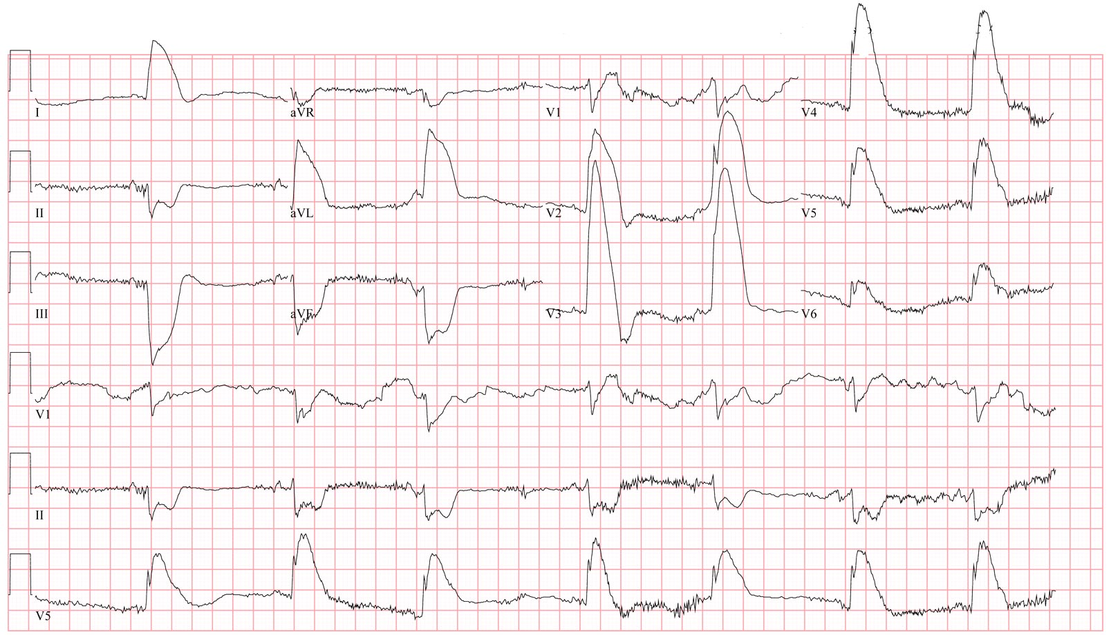
looks like a really really ugly ant lateral stemi.
get the iabp and vent ready, this guy is gonna be a sick puppy
Anony -
This is a really ugly anterior MI, but what makes it particularly ugly is how it happened: from an acute Type A (Debakey II) aortic dissection.
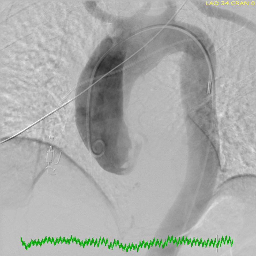
Note the huge ST segment elevation in the anterior precordial leads that exceed the height of the QRS complex. Because the patient presented with these EKG abnormalities and severe hypotension, the patient was taken emergently to the cath lab. A balloon pump was inserted and augmented the patient's blood pressure. A pigtail catheter was placed in the aorta and an aortic root shot performed. It shows the end of the pigtail catheter in the false lumen of the dissection that extends to the area just short of the arch vessels and no coronary arteries (they arise from the true lumen). Mild-moderate aortic insufficiency was noted at the same time, presenting a dilemma about the use of the balloon pump to support the patient's blood pressure. It was clear the dissection involved the left main coronary artery and aortic valve also.
The patient was rushed directly to the operating room and underwent and extensive repair, but ultimately did not survive to leave the hospital.
Post a Comment