Showing posts with label radiology. Show all posts

Friday, August 16, 2013

When Placing a Pacemaker, You Know You're on the Wrong Side When

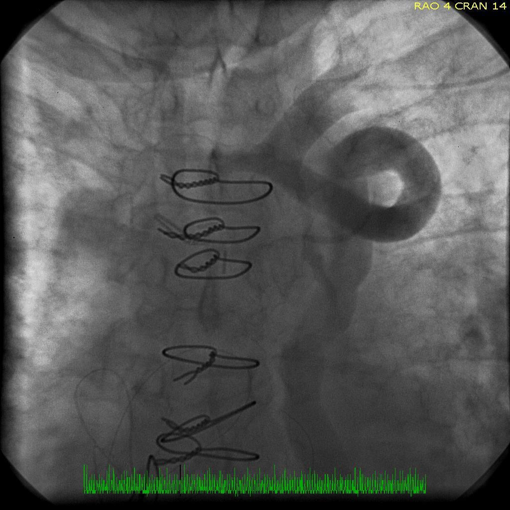
... your wires look like this: 

AP fluoroscopic view

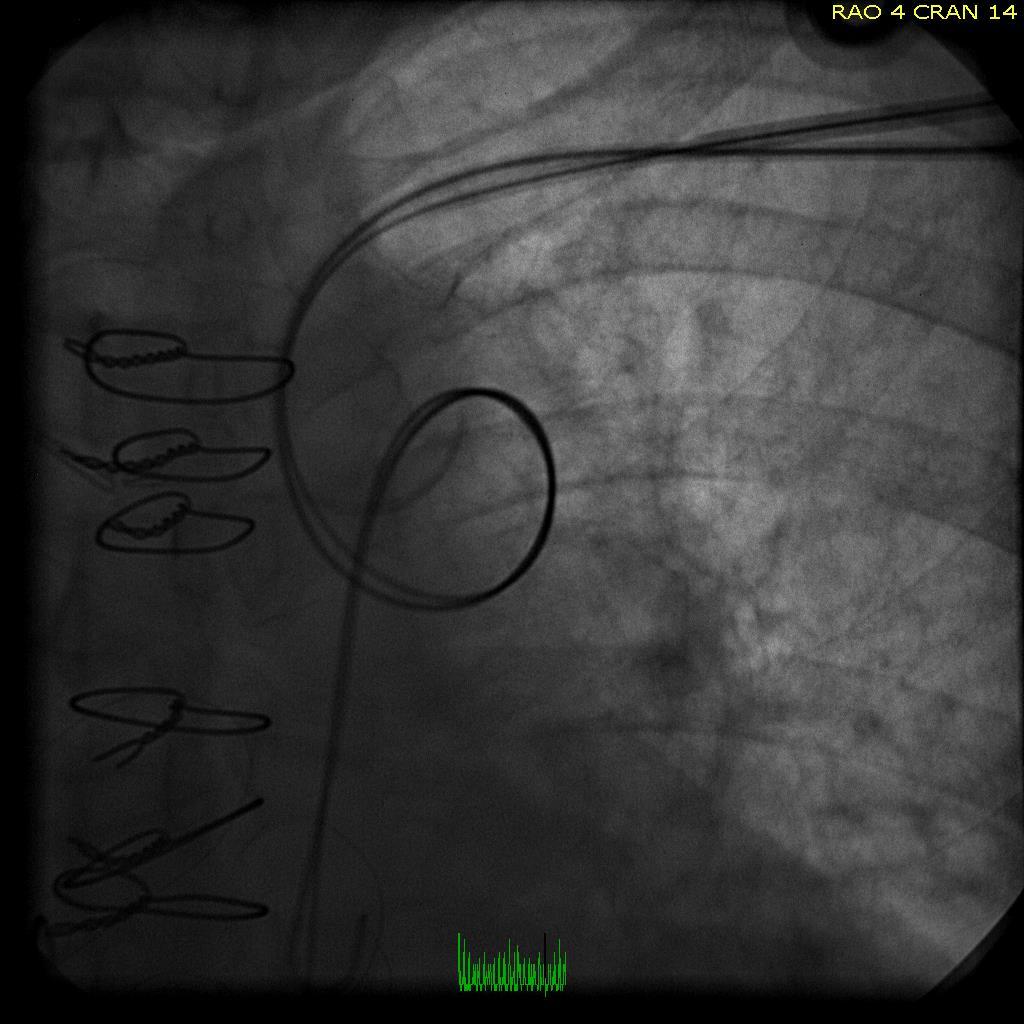
... and the venogram looks like this: 

Results of a left subclavian venogram

-Wes

Tuesday, July 02, 2013

A Chest X-Ray You Don't See Everyday

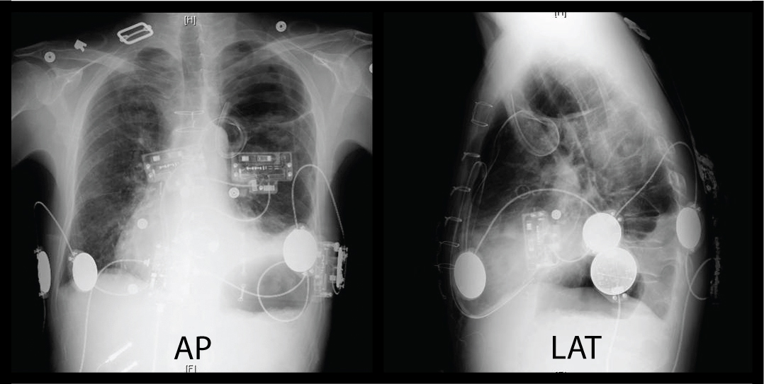
... unless, of course, you're a cardiac electrophysiologist:

An anterior-posterior (AP) and lateral (LAT) chest radiograph
(Click image to enlarge)
-Wes

PS: Answer here.

Sunday, February 24, 2013

How Big Data's Fueling Complacency

"What's the most important finding on this chest x-ray?"

There he was, standing before 5 ICU residents, each peering at a chest film on displayed on the over-sized computer screen.

"Um, the pleural effusion?" whimpered a third-year resident.

"No!" barked the attending.

The others, standing dumbfounded in front of the computer display, searching for another finding but finding none, stood silently.

"Come on, folks!  Look!"

And try as they may, no one saw it.

"The name, folks, the name!" the attending said impatiently.

And there it was, a tiny reminder of whose x-ray it was, quietly lurking in tiny print in the upper right corner of the computer screen, unmagnified. 

But wait, the name was correct.  What the heck was he talking about?

Closer inspection showed another critical piece of information, totally lost on almost everyone standing there: the date of birth of the patient.  It was not the same as the patient being discussed. They were looking at the wrong patient's chest x-ray.  Never mind that their patient had a chest tube placed on the opposite side that wasn't shown on the displayed chest x-ray.  Yet they were already trying to make decisions for care.

***

I recently taught an EKG reading class and had a similar experience to the one above.  Since July, I've been teaching the basics of EKG reading at least once a month: rate, rhythm, axis, intervals - you know the drill, right?

But I (once again) asked about the axis of an EKG tracing we were discussing some six months later.  A room full of at least twenty residents sat quietly.  No one answered.

I kept my composure.  I prodded them gently, hoping to hear an answer yet none came.  Were they on call?  Distracted by their cell phones or pending work?  Am I THAT boring?

Still nothing.

So I reviewed how we determine EKG axis, and quickly, a few remembered the concept and gratefully, responded correctly.

But these experiences got me thinking about the effects Big Data is having on our residents today and its tendency to build complacency.  Why learn something if you're always spoon-fed it right?

Admittedly, our medical data explosion has prevented us from knowing everything there is to know about anatomy, physiology, pathology, treatment options and the like.  There is a role for access to Big Data.

But increasingly the data we feed our residents and medical schools is nothing but printed characters: x-ray reports, EKG interpretations, study results like "ejection fraction:" all limited to the 256 ASCII character set.  Residents no longer feel the need to look at the raw image and formulate their own opinion - they'll just look at the printed report.  They expect the data to be fed to them in printed format.  They expect the reading to be correct.  In a way, they're growing up expecting to be spoon fed just the black-and-white answers rather than the brilliant data provided by pictures.  Just "google it."

Never mind the computer says "atrial fibrillation" because the original EKG contains noise.

Such an "Big Data-entitled" approach to health care is extremely dangerous, especially if the data upon which decisions are based, are wrong.  Residents should never forget two things my father always told me:

"Garbage in, garbage out" and "expect what you inspect."

-Wes
Wednesday, April 21, 2010

Will iPhone Apps Have to be FDA-Approved as Medical Devices?

... if you're a radiologist, it seems some do:

“We did a stroke trial ... and compared performance of radiologists reading on the iPhone to the standard clinical reading work station and the performance was identical,” said Mitchell. “They performed just as well in this tough diagnostic task.

“That’s a really good indicator that this could be quite useful for diagnosing all sorts of things that aren’t as tough,” he added.

Mitchell said it’s currently being tested on the new iPad, which has a larger screen to view the images.

The application for iPhone and other mobile devices has been licensed as a medical device in Canada, said Philippe Laroche, a spokesman for Health Canada.

It’s still awaiting approval from the US Food and Drug Administration and CE Mark in Europe.
Now, the next blockbuster: the FDA-approval iPhone app!

-Wes

Sunday, March 07, 2010

A Cardiology Website to Bookmark

Whether you're a medical student, resident, fellow, or staff cardiologist, once in a while you need a simple, succinct reference for a talk on the basics of cardiovascular disease. Who knew it would come from a radiologist:
LearningRadiology.com was conceived, designed, developed, is published, managed and maintained and its content is produced in its entirety by William Herring, MD, FACR. Dr. Herring is the Vice-Chairman and Radiology Residency Program Director at Albert Einstein Medical Center in Philadelphia, Pennsylvania, where he has been the Radiology Residency Program Director for over 25 years.

All material on the site, except for the Faculty lectures by other members of the staff at Einstein, was produced by Dr. Herring. Started in June of 2002, the site was originally intended to replace the handout notes that accompanied lectures for the residents and medical students at Albert Einstein Medical Center. It now contains over 20,000 pages of content, and has grown in popularity so that currently over 9 thousand unique visitors access the site every day. Visitors access over 20 million pages of content every year.

The site has been, and continues to be, free to use. It requires no registration, no sign-in, no fee and is commercial-free.
Kudos to Dr. Herring.

-Wes

h/t: Dr. S. Venkatesan, MD.

Friday, December 18, 2009

Beware of Voice Recognition Software for Reading Chest X-Rays

You might get something like this:
"Single view.

There are no prior studies for comparison. The heart is mildly enlarged. The ovarian veins are engorged area.

"The increased density in the medial portion of the right base may be parenchymal or due to vascular structures. The remainder of the lungs is air. No effusion."
Um, did I miss something in medical school?

Dang.

-Wes

Sunday, July 20, 2008

When Radiology Comes to Airport Security


... it can lead to all sorts of interesting findings:
The new full-body imaging machines that will arrive at O'Hare this fall look through clothing to create an explicit silhouette of the traveler—showing shapes, folds of fat and other anatomical characteristics—to identify possible hidden objects.

Even though facial features are blurred to protect privacy, the images reveal breasts, buttocks and other private parts, prompting some civil liberties groups to call the machines an unacceptable intrusion. ...

The full-body imaging pictures are formed by one of two ways—bouncing beams of radio frequency energy off the skin or conducting a low-dose X-ray of an individual.
Quick! Where's HIPAA?!? Imagine the breast implants, tummy tucks, pacemakers, defibrillators, mastectomy scars, colostomy bags, naval (and other appendage) jewelry, folds of fat, type of underwear they'll see. Worse yet, what if someone's going "commando" without underwear?!

Man, those poor airport security agents are going to need hazardous duty pay...

... oh, yeah, I forgot to mention...

... welcome to our world.

-Wes

Wednesday, April 30, 2008

The Wonders of Radiology Voice Recognition Software

It has become common practice for radiologists to migrate to voice-recognition software to expedite the processing of the myriad of reports that come their way and to save on transcription costs. Here is the actual final report received by one of our cardiologists and delightfully handed to me today:
"HISTORY: CHF.

COMPARISON: 04/20/2008.

Colon pulmonary vascular prominence is again identified, unchanged dirty mediastinal stool is enlarged, but stable. Atherosclerotic disease is present in the aortic arch. Bones are unremarkable.

IMPRESSION: Findings consistent with stable CHF.
XRAY CHEST SINGLE VIEW (Order #95511140) on 4/24/2008"
The radiologist was called and said, "What did I sign off on?"

We read back the report.

"Oh, my God!"

-Wes