Using wooden sticks, Niehoff and Evans touch the bars where they want Kwashi to place his hands. The gorilla complies.-Wes
"Good boy," Evans says, slipping grapes into the gorilla's mouth.
Evans applies conductive gel to the tip of a small wand that emits high-frequency sound waves. Then, with grapes in his left hand, Evans moves the wand in his right hand toward Kwashi's chest, being careful not to place the instrument completely through the bars.
This is where an important part of Kwashi's training kicks in. The gorilla leans forward slightly until his chest rests against the bars. He holds that pose - and continues gobbling grapes - as the wand touches his chest.
A few feet away, Jenny Schaaf stands at a state-of-the-art cardiac ultrasound machine on loan to the zoo from Toshiba. The monitor shows the gorilla's beating heart, which Schaaf, technical director of the echocardiography lab at Christ Hospital, captures for later analysis.
"Tilt up a little bit," she says, directing Evans on how to position the wand. "Little bit more. That's perfect."
"Good boy!" Evans says, as Kwashi holds his pose.
A few minutes later, though, Kwashi grunts. Evans recognizes the sound as the "cough vocalization."
"That's a warning that, 'I don't like what you're doing.' He doesn't like his chest being touched."
Showing posts with label echocardiography. Show all posts
Showing posts with label echocardiography. Show all posts
Tuesday, October 11, 2011
The Challenges of Gorilla Echocardiography
If you thought human echocardiography was tough, imagine what it's like to perform an echocardiogram on a gorilla while it's awake:


Thursday, June 17, 2010
Normal Echocardiogram Reading Error Rate 0.2% - By Technicians
In a "study" that is unlikely to ever be repeated again, we find that echocardiogram technicians are pretty good at screening normal echos, and have, according to credentialed cardiologists, a 0.2% reading error rate:
I wonder what the "normal" echocardiogram reading error rate is for board-certified cardiologists?
-Wes
Harlem Hospital Center has completed an investigation of 7,000 heart tests going back five years, most of which had never been reviewed by doctors, a spokeswoman for the hospital said on Wednesday.Recall that the echocardiogram technicians were only sending abnormal tests for cardiologists to review, and only presumed normal tests were never read (or signed) by a certified cardiologist.
The investigation found that 14 patients might have been misdiagnosed because their tests had not been handled properly, said the spokeswoman, Ana Marengo. Twelve of the patients had been contacted, and none of them were found to have suffered adverse effects from the failure to properly read their tests, Ms. Marengo said."
I wonder what the "normal" echocardiogram reading error rate is for board-certified cardiologists?
-Wes
Monday, February 01, 2010
The New Commodity on E-bay: Used Cardiac Ultrasound Probes
At nearly a $500-$1000 a pop when auctioned on E-bay, used phased-array cardiac ultrasound probes have recently become a hot commodity for theft in our local hospitals. This poses big problems when they're needed urgently in the electrophysiology or cardiac catheterization laboratories.
Be warned.
-Wes
Be warned.
-Wes
Monday, April 13, 2009
EchoJournal Launched
Call it YouTube for echocardiography. The guys over a Medgadget have launched EchoJournal:
I wish I had thought of that.
-Wes
PS: I'd be careful to make sure patient identifiers are removed from video loops before uploading, however.
It is a video site, designed to present transthoracic and transesophageal loops for discussion and storage. EchoJournal combines many of the features familiar to users of other video sharing sites, including easy uploading, comments and discussions, and the ability to create communities for your academic department, professional group, or school. The site will be curated by David E. Winchester, MD, a Cardiology Fellow at the University of Florida, who has already uploaded tons of interesting cases, and is ready for many more.Great idea.
I wish I had thought of that.
-Wes
PS: I'd be careful to make sure patient identifiers are removed from video loops before uploading, however.
Tuesday, January 27, 2009
Who Needs a Stethoscope?
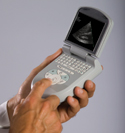
... when you can have a pocket-sized echocardiogram.
And better yet, this stethoscope comes with CPT procedure codes.
-Wes
Reference:
Specs on Seimen's Acuson P10 pocket-sized echo machine (pdf).
Monday, December 29, 2008
More Alphabet Soup
"ACC/AHA/ACR/ASE/ASNC/HRS/NASCI/RSNA/SAIP/SCAI/SCCT/SCMR/SIR 2008 Key Data Elements and Definitions for Cardiac Imaging"
I could not make those initials up. This was the title of the recently published paper in the Journal of the American College of Cardiology from the writing committee organized to develop clinical data standards for cardiac imaging.
Translation:
ACC = American College of Cardiology
AHA = American Heart Association
ACR = American College of Radiology
ASE = American Society of Echocardiography
ASNC = American Society of Nuclear Cardiology
HRS = Heart Rhythm Society
NASCI = North American Society for Cardiovascular Imaging
RSNA = Radiological Society of North America
SAIP = Society for Atherosclerosis Imaging and Prevention
SCAI = Society for Cardiovascular Angiography and Interventions
SCCT = Society of Cardiovascular Computed Tomography
SCMR = Society for Cardiovascular Magnetic Resonance
SIR = Society of Intervential Radiology
Wow, that's a tour-de-force of cardiac imaging specialists.* These are smart men and women who understand how the Medicare payment game ispaid played. They understand that data, lots and lots of data, in fact, so much data that your head will spin, is the way to assure Medicare payment. In short: gather lots and lots of data, any way you can, to assure the folks on the Hill that you are really serious about showing the cost effectiveness of all of this testing:
But tied to each of these data element sets created for every cardiac imaging test, is the requirement to include as an "administrative" data element the patient's insurance information. The writing group explains this data-element necessity as a means to "foster optimal cardiovascular care and disease prevention" because:
Imagine, line item data on your renal function, ejection fraction, stress test results, cardiac risk factor analysis - all instantly available to an insurance company's computer database and conveniently entered unwittingly by the echocardiography clerical staff in the hospital of your choice. Imagine the next time you then go to purchase insurance. Best of luck to you.
Or imagine you are a doctor without an electronic medical record and ordering system - best of luck to you as you complete the test-request form that will soon look more formidable than the MCAT bubble answer sheet used to qualify for medical school. Remember to use a number 2 pencil.
Or consider if a clerical error is made during data entry: what recourse will you have? Will you have the option of editing the data entered to assure insurer payment for the test they claim was not appropriate because four data fields were left blank? "Sorry Mr. Jones, on the basis of a creatinine of 1.6, and four empty data fields on the ordering questionnaire, this test should not have been ordered, so * b-o-o-i-i-n-n-g-g *, we're not going to pay for that test after all."
But in this era of "evidenced-based medicine," these convoluted, complicated, and thoughtfully-produced-but-arbitrary data forms (Note: 20 pages were devoted in the article just for the medical history fields alone. And what is an "equivocal" test anyway?) are being feverishly developed by these cardiac imaging societies. They must justify continued testing and Medicare payments on the basis of a promise of future as-yet-undeveloped studies based on retrospective utilization data completed by the non-medical clerks at the time of test ordering. And thanks to hoards of well-meaning academics that are eager to secure more grant funding, this same dubious retrospective data will soon serve as paradigms upon which our treatment and further testing "guidelines" will be based.
But retrospective data collection like this is far easier and less expensive to gather rather than a prospective, randomized trial. Who needs those?
Heck, I guess I should stop complaining.
After all, I am a cardiologist.
-Wes
Reference:
"ACC/AHA/ACR/ASE/ASNC/HRS/NASCI/RSNA/SAIP/SCAI/SCCT/SCMR/SIR 2008 Key Data Elements and Definitions for Cardiac Imaging." J Am Coll Cardiol, 2009; 53:91-124, doi:10.1016/j.jacc.2008.09.006 (Published online 8 December 2008).
I could not make those initials up. This was the title of the recently published paper in the Journal of the American College of Cardiology from the writing committee organized to develop clinical data standards for cardiac imaging.
Translation:
ACC = American College of Cardiology
AHA = American Heart Association
ACR = American College of Radiology
ASE = American Society of Echocardiography
ASNC = American Society of Nuclear Cardiology
HRS = Heart Rhythm Society
NASCI = North American Society for Cardiovascular Imaging
RSNA = Radiological Society of North America
SAIP = Society for Atherosclerosis Imaging and Prevention
SCAI = Society for Cardiovascular Angiography and Interventions
SCCT = Society of Cardiovascular Computed Tomography
SCMR = Society for Cardiovascular Magnetic Resonance
SIR = Society of Intervential Radiology
Wow, that's a tour-de-force of cardiac imaging specialists.* These are smart men and women who understand how the Medicare payment game is
The ACC and AHA recognize the importance of the use of clinical data standards for patient management, to assess outcomes, and conduct research, and the importance of defining the processes and outcomes of clinical care, whether in randomized trials, observational studies, registries, or quality improvement initiatives. Hence, clinical data standards strive to define and standardize data relevant to clinical topics in cardiology, with the primary goal of assisting data collection by providing a platform of data elements and definitions applicable to various conditions. Broad agreement on a common vocabulary with reliable definitions used by all is vital to pool and/or compare data across studies and assess the applicability of research to clinical practice. The growing adoption of electronic medical records renders an even more imperative and urgent need for such definitions and standards. Therefore, the ACC and AHA have undertaken the task of defining and disseminating clinical data standards—sets of standardized data elements and corresponding definitions to collect data relevant to cardiovascular conditions. The ultimate purpose of clinical data standards is to contribute to the infrastructure necessary for accomplishing the ACC/AHA’s mission of fostering optimal cardiovascular care and disease prevention.On the surface, these efforts seem like the best way to begin to get a handle on the cost-effectiveness of cardiac testing. No doubt the shear volume of data generated will be heretofore unmatched and a rich source of research grants for academe for years to come.
But tied to each of these data element sets created for every cardiac imaging test, is the requirement to include as an "administrative" data element the patient's insurance information. The writing group explains this data-element necessity as a means to "foster optimal cardiovascular care and disease prevention" because:
The insurance payer element was included to be certain that patients of all payer status were included in studies equitably, especially those funded federally (ed: emphasis mine). The inclusion of this data was not to in any way suggest that cardiac imaging patients should be screened on the basis of ability to pay.Yep, it's all to justify to the Medicare National Bank that, yes sir, we really DID need this test. While the test might be ordered irrespective of the ability of the patient to pay as the writing committee suggests, for the first time we will be granting insurers direct automated line-item access to clinical information and the test results. The implications of this are concerning.
Imagine, line item data on your renal function, ejection fraction, stress test results, cardiac risk factor analysis - all instantly available to an insurance company's computer database and conveniently entered unwittingly by the echocardiography clerical staff in the hospital of your choice. Imagine the next time you then go to purchase insurance. Best of luck to you.
Or imagine you are a doctor without an electronic medical record and ordering system - best of luck to you as you complete the test-request form that will soon look more formidable than the MCAT bubble answer sheet used to qualify for medical school. Remember to use a number 2 pencil.
Or consider if a clerical error is made during data entry: what recourse will you have? Will you have the option of editing the data entered to assure insurer payment for the test they claim was not appropriate because four data fields were left blank? "Sorry Mr. Jones, on the basis of a creatinine of 1.6, and four empty data fields on the ordering questionnaire, this test should not have been ordered, so * b-o-o-i-i-n-n-g-g *, we're not going to pay for that test after all."
But in this era of "evidenced-based medicine," these convoluted, complicated, and thoughtfully-produced-but-arbitrary data forms (Note: 20 pages were devoted in the article just for the medical history fields alone. And what is an "equivocal" test anyway?) are being feverishly developed by these cardiac imaging societies. They must justify continued testing and Medicare payments on the basis of a promise of future as-yet-undeveloped studies based on retrospective utilization data completed by the non-medical clerks at the time of test ordering. And thanks to hoards of well-meaning academics that are eager to secure more grant funding, this same dubious retrospective data will soon serve as paradigms upon which our treatment and further testing "guidelines" will be based.
But retrospective data collection like this is far easier and less expensive to gather rather than a prospective, randomized trial. Who needs those?
Heck, I guess I should stop complaining.
After all, I am a cardiologist.
-Wes
* The conflicts of interest of the authors of this document warrant notice. They are not insignificant - so much so that the article reviewers conflicts were also disclosed as an appendix to reassure a more "non-biased" critical review of paper occurred.
Reference:
"ACC/AHA/ACR/ASE/ASNC/HRS/NASCI/RSNA/SAIP/SCAI/SCCT/SCMR/SIR 2008 Key Data Elements and Definitions for Cardiac Imaging." J Am Coll Cardiol, 2009; 53:91-124, doi:10.1016/j.jacc.2008.09.006 (Published online 8 December 2008).
Friday, February 01, 2008
Imaging Turf Wars
Well the inaugural issue of JACC Cardiovascular Imaging was just come out and heralds cardiologists as cardiovascular imaging experts. Eugene Braunwald's welcome letter was telling:
Why?
It's all about the money.
-Wes
"Despite its great advantages, selective angiography is an invasive technique that may be uncomfortable, entails some minimal risk to the patient, and is expensive. It can be applied only a limited number of times in each patient and it is not suitable for screening. Therefore, physicians caring for patients with known or suspected heart disease yearned for noninvasive cardiac imaging. Fortunately, 2 new techniques—echocardiography and nuclear imaging—became available in the 1960s and have improved progressively since then. Literally tens of millions of these examinations are performed each year, transforming medical practice. In the 1980s, 3 new noninvasive imaging techniques—computed tomography (CT), cardiac magnetic resonance (CMR) imaging, and positron emission tomography (PET, a nuclear technique)—emerged. Each of these provides information on cardiac structure and function that is an order of magnitude greater than the earlier imaging techniques.Radiologists are none too happy, I'm sure. No doubt they might take issue with the claim of "We need a new generation of cardiac imagers who are expert with the entire portfolio of modalities and who can provide a nonbiased selection of the technique that can best solve the clinical or research problem at hand." The friendly collaboration between radiologists and cardiologists went out the window long ago.
None of the aforementioned techniques are static and all are undergoing progressive refinement. Many important advances have been made by subspecialists in a single technique such as echocardiographers, nuclear cardiologists, and specialists in CT, CMR, or PET imaging, respectively. Each of these imaging modalities have their individual training programs, and the advances in these fields are described in specialized journals, each devoted to a single technique. Although this approach has been responsible for spectacular advances in each modality, it has led to fragmentation of patient evaluation, at times to competition between individual modalities, and to increased costs at a time of diminishing resources to pay for health care.
There is a growing consensus that the time has come to consider cardiovascular imaging in a more comprehensive, unified manner. We need a new generation of cardiac imagers who are expert with the entire portfolio of modalities and who can provide a nonbiased selection of the technique that can best solve the clinical or research problem at hand. To develop these broadly-based experts, we need new unified training programs that break down what are sometimes artificial interdepartmental and intradepartmental barriers.
Another important tool in shifting the imaging paradigm is to develop a journal of cardiovascular imaging. The Publications Committee of the American College of Cardiology and its talented staff, working closely with the College leadership, developed a vision for and then the concrete plans for the creation of JACC: Cardiovascular Imaging. The committee and its consultants selected the Editor-in-Chief, Dr. Jagat Narula, from a group of distinguished cardiac imagers. Dr. Narula has put together an outstanding group of associate editors, editorial board members, and consultants. They now deserve the cooperation of the entire cardiovascular community as they proceed on their important mission."
Why?
It's all about the money.
-Wes
Sunday, October 07, 2007
Echocardiogram Contrast Agents to Get Black Box Warning
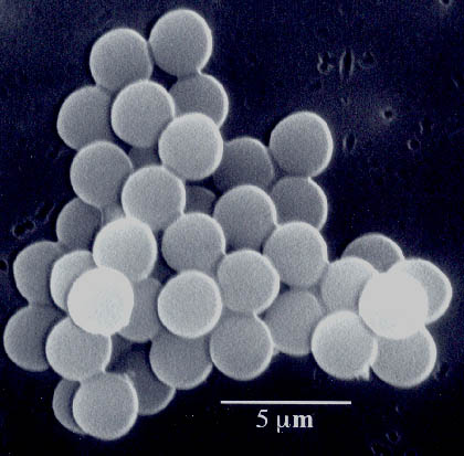 It seems the FDA is about to issue a block box warning on echocardiographic contrast agents:
A spokeswoman for the agency told the Health Blog that it “has been investigating reports of deaths and serious cardiopulmonary reactions.” The FDA will alert health-care professionals about the “serious risks” and “provide guidance regarding the safe use” of the drugs, she said. The warning could come later this week.It seems that these agents have been associated with some extention of the EKG's QT interval slightly and there are concerns they may be proarrhythmic. These agents already have a warning regarding their use in patient's with suspected cardiac shunts because the phospholipid-encapsulated microspheres can bypass the lung's natural filtering mechanism and potentially cause microsphere trapping in small arterioles under 15 mm in size, especially at branch points. it is interesting to note that deaths have been reported late after the use of these agents:
The contrast agents are used in a small fraction of echocardiograms, the FDA said. Doctors often turn to the drugs when ultrasound without them is inconclusive.
The principal drug at issue is Definity, made by Bristol-Myers Squibb. A second drug called Optison, from General Electric, is also expected to get the same labeling treatment. The drugs contain microscopic gas-filled spheres and are injected into the veins of patients to punch up the contrast of images for tough cases. After a few minutes the gas leaves the body.
The agency has asked the manufacturers to update their product labeling, which they’ve agreed to do, the FDA spokeswoman said. So look for some new black boxes, the sternest warnings available, that would tell doctors not to use the medicines in patients with unstable angina, acute heart attacks and unstable cardiopulmonary disease, according to people familiar with the matter.
It’s also expected that the labels would instruct doctors to monitor patients’ heartbeats for trouble with an electrocardiogram, or ECG, for a half-hour after the ultrasound exam is finished.
Among the 1716 activated DEFINITY® patients, 19 (1.1%) suffered serious cardiopulmonary adverse events including eight deaths. The deaths occurred several days after activated DEFINITY® administration and appear to be related to the course of underlying disease. Of the 11 other serious adverse events, which appeared within days of the drug administration (2-15 days), all appeared to be a progression underlying cardiac and non-cardiac disease.These imaging agents can really help evaluate the heart function of our increasingly obese population undergoing echocardiography, but the logistical nightmare required to monitor these patients after their echo might significantly limit their use.
I'll update this post when the FDA's statement is issued.
-Wes
Reference: Image of microspheres from Kenneth S. Suslick, University of Illinois, Urbana-Champaign.
Wednesday, April 11, 2007
The Hand-Carried Cardiac Ultrasound
Perhaps a useful tool in the evaluation of murmurs and heart muscle function in the cardiology and medical clinic settings, the hand-carried cardiac ultrasound device might reduce the need for conventional echocardiography. But given that cardiologists earn significant income from performing and interpreting standard echocardiograms, would they ever use this device?
-Wes
-Wes
Subscribe to:
Posts (Atom)