The following is an actual cardiac electrophysiology case study offered with the patient's permission. It's technical and contains an image that might turn some folks' stomachs, so for those who are a bit squeemish or just ate a meal: consider yourself warned and feel free to pass on this post. For the rest of you who remain interested and don't mind medical images, good luck.
It was supposed to be a simple ICD revision.
A prior abdominal ICD has been implanted in 1995. As was the norm of the day, the large ICD pulse generator was implanted in the left upper quadrant of the abdomen and an connected to an old Guidant Endotak Model 0074 lead that was implanted via the left subclavian vein and then tunneled down to the abdominal pocket. The device served the patient well for many years until its battery depleted in 2003. At that time, a new, smaller ICD with an appropriate header replaced the old abdominal device and because the defibrillator lead worked well, the smaller ICD pulse generator was left in the abdominal pocket.
Years passed and the patient followed reliably in the Device Clinic for his routine defibrillator checks. While the lead impedance and capture thresholds remained normal, about a year ago intermittent periods of noise with non-physiologic short RR intervals suggestive of possible impending lead fracture began to appear on the patient's device checks. Because the patient was not pacemaker dependent nor near the time when his existing ICD battery would have to be replaced again, it was elected to wait until his battery reached it's elective replacement indicator before revising his system. When that time came, a new defibrillator lead could be implanted and connected to a more conventional VVIR ICD pulse generator implanted in the upper chest area (the patient has chronic atrial fibrillation). The old pulse generator could then be removed from his abdomen and the old lead capped and left in place.
So the day came for surgery. The patient felt fine: no fever, chills or other unusual symptoms pre-operatively. A venogram performed immediately before the procedure disclosed a patent left axillary and subclavian veins, so it was decided to first proceed with the new ICD implant on the same side as the site where his first defibrillator was implanted followed by removal of the old ICD pulse generator from the abdomen. Pre-operative antibiotics were administered. To make a long story quite a bit shorter, the new single-chamber ICD implanted via the left axillary approach was performed without a hitch. A dressing was applied to the wound and preparations made to explant the abdominal pulse generator.
The lower abdominal area was similarly prepped and draped. Local anesthetic was infiltrated over the prior abdominal scan and an incision made at this location. Using electrocautery dissection, the incision was carried to the pulse generator capsule which appeared to be quite thick, but uninflammed. The fibrous capsule surrounding the pulse generator was then opened. What was found was startling to all.
Inside the pulse generator pocket was the device and lead system surrounded in a thick fluid that looked, for lack of a better way to describe it, like wet, brown mud. There was no odor. The device was extracted from the pocket after the suture holding the header of the device to the pocket wall was cut. A portion of the lead was also cut removed with the device. A picture of the removed device is shown here:
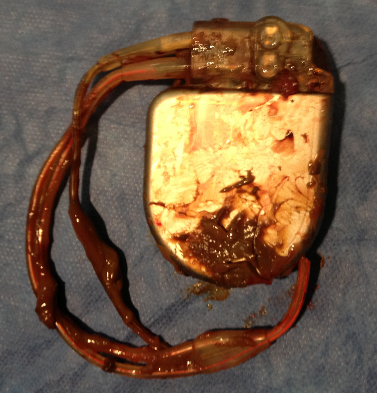
Soooo. What now?
Imagine you are the surgeon with this device in your hands. You have another case after this one. You struggle to find where this situation falls within our clinical "guidelines" for care and find very little. You aren't sure what you're seeing, but only know that this "chocolate-coated" ICD pulse generator is not the norm. (Usually they are nice and clean without debris.)
Ideas?
-Wes
3 comments:
Wes,
I've come across this exact situation 3 or 4 times before and am pretty sure I know what is going on. We have seen this in second or later pulse generator changes and more commonly in abdominal pockets.
When an ICD is replaced into a chronic ICD pocket, the remaining open space in the pocket can serve as a cavity that can collect debris. If pocket capsule is not dissected away partially or completely, the closed pocket will act as a protected closed space. Any remaining blood from the prior surgery will have no place to go. As the generator rubs in the interior of the capsule, the capsule material will slough off and collect in the pocket. Because there is no communication with the subcutaneous space, the debris will simply fill the additional space. The debris is not infectious.
This is not seen at the time of first pulse generator changes, because the pocket forms around the new device as the remaining surgical blood drains away. I suspect it happens less often in pectoral pockets, because typically we have to dissect the pocket at least a bit to fit the new device in place. I think any form of extrapocket communication with the subcu tissue will prevent the process. In addition, these devices don't generally get banged around as much as the abdominal devices.
I first heard about type of case this from Rick Henthorn, your old partner. He was concerned about infection and found none. I think he got surgical consultation and the pocket was debrided. Later when I saw the same process on another abdominal changeout, I was ready for it and treated it conservatively.
Since then, I never close a chronic device pocket without at least some form or capsulotomy. So far, I have not seen this happen again.
For your patient, my approach would be to wash out all the debris and carve out a portion of the abdominal pocket to allow communication with the subcutaneous space. I'd send the debris for culture, but would not expect it to be positive. Personally, I'd hold off on further antibiotics unless the cultures were positive.
Great case. Please share your thoughts and findings.
Jay
@EJSMD
Jay -
Ah yes. It always helps to have a veteran reading your blog.
As you say, this is precisely what was found. Gram stain of the debris showed no white cells or organisms. The capsule was removed en its entirety. Cultures of the pocket capsule tissue, blood cultures, and cultures of the fluid all came back no growth. The patient had been on warfarin (and later Xarelto) for his atrial fibrillation.
Of course, at the time of the procedure, the culture information was not known. So the new ICD pocket was re-draped, prepped, and the wound opened to find the tunneled 0074 ICD lead's tie-down sleeve. This was released and the lead cut, capped, and the distal portion withdrawn back to the abdominal pocket to prevent a "wick" extending from the abdominal wound to the new ICD system. The new ICD wound was re-closed.
The addominal would was irrigated with antibiotic saline solution, closed primarily, and a Jackson-Pratt drain placed to remove deadspace so no seroma formation could occur. The antibiotic was expanded to Vancomycin only (from Ancef) and continued for three days then disconinued when the Jackson-Pratt drain was removed.
The patient was seen by ID who agreed that stopping antibiotics and observing was the best scenario. He was seen in follow-up 10 days later and has done well.
This residue looks like the contents of an endometrioma. It is frequently called a chocolate cyst given how food oriented much of doctor terminology is. It is also the result of blood breakdown and is likewise sterile.
Post a Comment